
- ELISA 클리아 FIA
- 핵산 추출 핵산 추출 및 PCR 키트 실시간 PCR
- 혈전 분석 혈액 그룹화 혈소판 집계
- POCT-FIA 콜로이드 골드 혈액 포도당 콜레스테롤 SARS-CoV-2
- 네오 ECG 홀터 모니터 휴대용 ECG 모니터 AI-ECG 포켓 ECG
- 환자 모니터 중요한 징후 모니터 엔드 조석 Capnography
- 핑거팁 산화계 휴대용 산화계 손목 산화계 웨어러블 산화계 올인원 건강 모니터 POCT 솔루션
- OBS 잠금 압축 플레이트 잠금 판 기존 플레이트 미니 플레이트 시스템 나사 Patella 반지-특허 제품 금속 핀 케이블 시스템
- 내부 고정 시스템 Laminoplasty 플레이트 시스템 경피 Kyphoplasty 퓨전 케이지 자궁 경부 고정 시스템
- 엉덩이 시스템 무릎 시스템
- 봉합 앵커 봉합 버튼
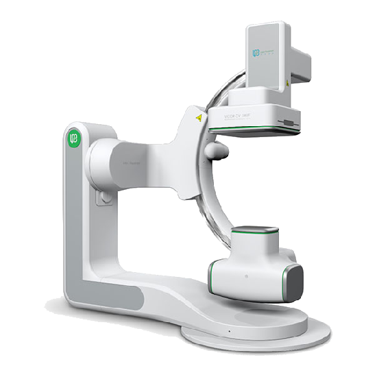
Vicor SWIFT 의료 혈관 X-ray 시스템 특징
1 고화질 및 안정적인 이미지
임상 적으로 정확하고 효율적인 진단을 지원하는 고해상도 및 와이드 필드 이미지
◆Digital flat panel detector
◆ Multiple panel models
◆ Adjustable, and wide-field
◆ Panel-digitized dynamic range up to 16 bit
◆ Accurate and clear details
◆ 1956 × 1956 까지 해상도
2 Long-lasting and Efficient Performance
◆3.0M heat capacity X-Ray tube, having long-lasting and stable life.
◆ Liquid-metal lubricated bearing to improve heat dissipation efficiency.
◆ 트리플 포커스 스팟, 다른 임상 응용 프로그램에 대한 명확한 이미지를 제공합니다.
3 안전한 방사선 보호
◆ Accurate working of the collimator to effectively reduce scattered rays
◆ 방사선량에 대한 효과적인 감독을 실현하기 위해 작동 중 RDSR로 능동 방사선량 관리 및 모니터링 및 방사선 선량 및 기타 정보의 실시간 및 정확한 기록
◆ Function of virtual collimator, to locate the checked site without X-ray egression
◆ 다양한 요구를 충족시키고 저선량으로도 고품질 이미지를 얻는 다중 용량 모델
4 전문 소프트웨어 시스템
Vicor CV Workstation
◆ 64-bit operating system, providing user-friendly interactive interface
◆ Accelerated real-time processing module with efficient GPU, and stable and reliable image
◆ Supporting multiple image processing tools such as automatic measurement, ventricular analysis and QCA analysis
◆ Real-time function of virtual beam limiter, helpful to reduce scattered rays and improve the safety of both physicians and patients
◆ Radiation dose report in compliance with IEC standard
◆ 네트워크 안전 표준을 준수하는 안전 설계
Vicor AngioExpert
◆ DICOM3.0 및 관련 표준을 준수하는 데이터 전송
◆ Supporting data storage and migration management
◆ Efficient completion of vascular contour and morphological data analysis, and generation of QCA analysis report
◆ Supporting dynamic correction of stent position to enhance stent visualization
◆ 완전 자동 원활한 이미지 접합 진단 범위를 확장하고 사지 세부 사항을 명확하게 표시
5 모든 부서에서 임상 응용

6 스텐트 향상
As an image recognition technique, it is used to realize the motion compensation for the X-ray image to clearly and accurately display the fine structure of the stent, and help physicians immediately evaluate the release of the stent during the operation.
* 이 기능은 판매와 상세한 상담이 필요합니다
7 스위프트 3D
완벽한 3D 재구성 기능을 통해 Vicor-CV SWIFT는 볼륨 재구성 (VR), 다중 평면 재구성 (MPR), 최대 강도 투영 (MIP) 및 최소 강도 투영 (MINIP) 과 같은 재구성 기술을 지원합니다. 혈관의 3 차원 형태와 공간적 관계를 분석하는 데 큰 이점이 있으며 혈관 시스템 질환, 종양 등에 대한 중재 적 치료에서 큰 진단 적 가치가 있습니다.
1. 혈관 이미지를 더 선명하고 정확하게 만들기 위해 혈관 구조가 겹치는 것을 피할 수 있습니다.
2. 병변 부위와 다른 정상 조직 및 기관 사이의 3 차원 관계 및 관강 구조는 어떤 각도에서도 관찰 될 수 있습니다.
3. 조영제의 복용량, 진단 및 치료 및 수술 시간 및 노출 용량을 줄일 수 있습니다.
4. 정확한 측정 결과는 수술 및 중재 치료를위한 데이터를 제공 할 수 있습니다
Lepu Medical에 관심을 가져 주셔서 감사합니다!
질문이나 문의를 이메일로 보내거나 연락처 데이터를 사용하십시오. 우리는 당신의 대답에 기쁠 것입니다 질문.